
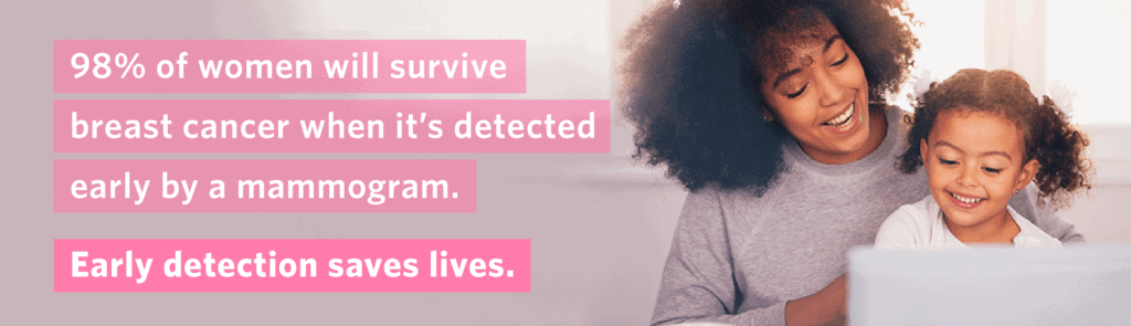
Your Mammogram is Important
To make sure you get the screening you need, we offer evening and weekend hours. Your mammogram is important. Take care of yourself and book yours today.
Jefferson Radiology offers the most advanced 3D Mammography technology available on the market today. This technology, called Genius™ mammography is offered at each of our locations.
Our exams provide better, earlier breast cancer detection compared to 2D alone and detect 20-65% more cancer. In fact, as many as 20% of breast cancers will be missed by 2D mammography. Greater accuracy means better breast cancer detection and a reduced chance of additional screenings.

Jefferson Radiology is recognized as a Mammography Accredited Facility by the American College of Radiology.
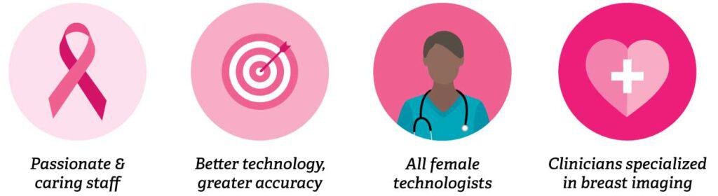
We’re revolutionizing mammograms with state-of-the-art 3D technology that offers shorter appointments, with scan times lasting just seconds. Our compassionate technologists are with you every step of the way, ensuring you get the peace of mind you deserve with accurate mammograms you can trust.
Book Your Appointment: 860-291-6569
A Genius™ 3D exam provides our radiologists with a series of detailed images of the breast. This allows them to better evaluate breast tissue layer by layer, making fine details more visible and no longer hidden by overlapping tissue.
The process of a Genius™ 3D mammography exam is the same as your conventional 2D exam. The technologist will position you, compress your breast, and take images from different angles. There’s no additional compression required.
The technologist will view the images of your breasts at the computer workstation to ensure quality images have been captured for review. A radiologist will then examine the images and report results to either your physician or directly to you.
As of January 1st 2019, 3D mammography screening exams are covered by Medicare and private insurers in the state of Connecticut and not subject to copays.
*Screening mammograms are considered a preventative screening exam and as such, are not subject to co-pays.
The Genius™ 3D exam has been clinically proven to greatly reduce the chance of callbacks, which are common for women with dense breasts. It is the only FDA approved 3D mammography machine to offer superior performance in women with dense breasts for routine breast cancer screening over standard 2D mammography.
A screening mammogram is generally covered as a yearly preventative exam through your insurance.
Based on your clinical history and your referring physician’s order, you may need a diagnostic mammogram. This type of mammogram requires more detailed images from different angles that allow the radiologist to check the area more closely. A diagnostic mammogram is applied toward the patient’s deductible.
Breast Ultrasound: this non-invasive exam provides a better view of the breast, cysts, or masses so your radiologist can provide a more accurate diagnosis.
Breast MRI: this exam provides more information after an abnormal mammogram. It’s considered more invasive since contrast is given through an IV before the procedure.
Breast Biopsy: this procedure takes cells/tissue samples with abnormal findings from the area.